
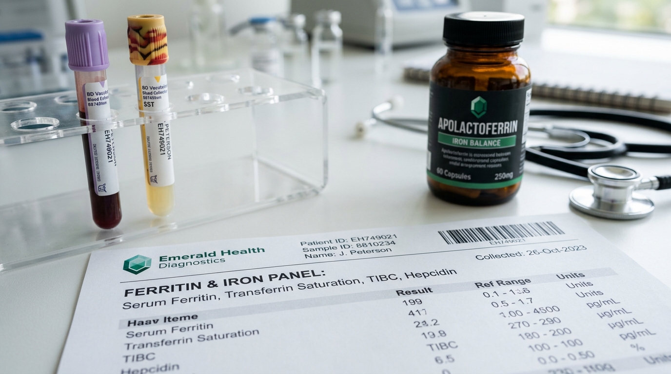
Evaluating iron metabolism through standard serum iron or basic hemoglobin panels alone is one of the most pervasive diagnostic failures in modern medicine. A patient can exhibit normal hemoglobin while suffering from profound, debilitating cellular iron deficiency, or conversely, harbor toxic, tissue-destroying iron overload while routine bloodwork appears unremarkable. In 2026, rigorous clinical biochemistry mandates evaluating the complete iron diagnostic triad: Serum Ferritin (intracellular iron storage), Transferrin Saturation (TSAT) (iron traveling in plasma), and Total Iron Binding Capacity (TIBC). Balancing this delicate equilibrium requires mastering Hepcidin regulation and deploying targeted interventions like Apolactoferrin rather than antiquated, gut-destructive ferrous sulfate pills.
- The Optimal Ferritin Window: While standard laboratory reference ranges define “normal” ferritin as 15 to 300 ng/mL, optimal cellular bioenergetics and hair follicle retention require 50 to 100 ng/mL for women and 75 to 150 ng/mL for men. Ferritin under 30 ng/mL triggers severe mitochondrial ATP depletion.
- The Fenton Reaction Hazard: When Serum Ferritin breaches 250 ng/mL alongside a Transferrin Saturation exceeding 45%, unbound free iron catalyzes the generation of hydroxyl free radicals via the Fenton Reaction, inducing lipid peroxidation in the liver, pancreas, and coronary endothelium.
- The Hepcidin Inflammatory Block: Elevated systemic inflammation (high hs-CRP or IL-6) triggers hepatic production of hepcidin, the master iron-regulatory hormone. Hepcidin degrades ferroportin channels, trapping iron inside macrophages and mimicking iron deficiency in bloodwork while tissues are simultaneously overloaded.
Clinical Iron Diagnostic Matrix: Differential Bloodwork Patterns
Interpreting iron panels requires evaluating Ferritin, Transferrin Saturation, and inflammatory markers concurrently:
| Clinical Condition | Serum Ferritin | Transferrin Saturation (TSAT) | TIBC (Binding Capacity) | Underlying Pathophysiology |
|---|---|---|---|---|
| True Absolute Iron Deficiency | < 30 ng/mL (Depleted) | < 16% (Severe Deficit) | High (> 400 mcg/dL) | Depleted bone marrow stores; insufficient iron for erythropoiesis |
| Anemia of Chronic Disease (ACD) | Normal to High (150–400) | < 20% (Functional Starvation) | Low to Normal (< 300) | High hepcidin blocks iron release; iron trapped inside reticuloendothelial cells |
| Optimal Healthspan Target | 70 – 125 ng/mL | 25% – 38% | 300 – 360 mcg/dL | Maximum mitochondrial cytochrome function with zero oxidative stress |
| Hereditary Hemochromatosis (C282Y) | > 400 – 1,200+ ng/mL | > 50% – 90% (Critical) | Low (< 250 mcg/dL) | Failure of hepcidin synthesis; uncontrolled gastrointestinal iron absorption |
The Hepcidin Regulatory Trap: Why Ferritin is an Acute-Phase Reactant
Serum Ferritin is not merely an iron storage vessel; it is an acute-phase inflammatory reactant. In the presence of systemic low-grade vascular inflammation (detectable via high-sensitivity C-reactive protein), the liver dramatically accelerates ferritin synthesis regardless of actual iron stores.
For therapeutic strategies targeting chronic endothelial inflammation, inspect our clinical protocol on hs-CRP & Systemic Vascular Inflammation: Curcumin Phytosome & Boswellia. The molecular mechanism operates via the IL-6 / STAT3 pathway:
- Circulating inflammatory cytokines stimulate hepatic hepatocytes to overproduce Hepcidin.
- Hepcidin binds to Ferroportin, the sole known iron-export protein on duodenal enterocytes and splenic macrophages.
- The hepcidin-ferroportin complex is internalized and degraded in lysosomes, halting iron export into plasma.
- Result: The patient’s red blood cell precursors are starved of iron (low TSAT), causing severe fatigue, while ferritin shows a deceptively elevated reading of 200+ ng/mL. Prescribing iron supplements in this scenario fuels systemic microbial growth without improving tissue oxygenation.
Apolactoferrin vs. Ferrous Sulfate: The Bioavailability Revolution
Standard oral iron therapy (ferrous sulfate 325mg, delivering 65mg elemental iron) has an abysmal compliance rate exceeding 60% due to severe constipation, nausea, and abdominal cramping. More dangerously, unabsorbed inorganic iron reaches the colon, triggering bacterial dysbiosis and mucosal inflammation.
Apolactoferrin (iron-depleted bovine lactoferrin) represents a breakthrough in therapeutic iron delivery:
- Natural Receptor Binding: Lactoferrin binds iron with 300x higher affinity than transferrin and is recognized by specific lactoferrin receptors in the brush border membrane of the small intestine.
- Direct Ferroportin Modulation: In head-to-head clinical trials, 200mg–250mg of Apolactoferrin restored serum ferritin and hemoglobin as effectively as 156mg of ferrous sulfate, while decreasing serum IL-6 and downregulating hepcidin.
- Zero Free Radical Burden: Because iron remains securely chelated within the lactoferrin glycoprotein pocket, it cannot participate in the destructive Fenton reaction inside the gut lumen.
Clinical Iron Restoration Protocol (For Ferritin < 40 ng/mL)
For individuals presenting with true iron deficiency without chronic inflammation:
# The Clinical Iron Restoration Blueprint
Morning (Fasting / 30 mins before breakfast):
-> Apolactoferrin: 250 mg
-> Iron Bisglycinate (Chelated): 25 mg – 36 mg elemental iron
-> Liposomal Vitamin C: 500 mg (Enhances duodenal DMT1 iron uptake)
-> Copper Bisglycinate: 1 mg – 2 mg (Essential for Ceruloplasmin ferroxidase activity)

Absorption Inhibitor Guardrails:
-> Zero coffee, black tea, or dairy for 2 hours post-administration (tannins and calcium block absorption by up to 80%).
-> Alternate-day dosing protocol: Taking iron every other day reduces hepcidin spikes by 45% compared to daily dosing.
For individuals with impaired metabolic clearing or elevated blood glucose, integrate this with our diagnostic guide on Fasting Insulin & HOMA-IR vs. HbA1c in Early Metabolic Dysfunction.
Iron Overload Defense: Phlebotomy & Phytate Chelation
In men and postmenopausal women carrying HFE gene mutations (hemochromatosis) or consuming excess red meat, iron accumulates relentlessly. Free iron deposits in cardiac myocytes, inducing arrhythmias and coronary artery disease. Therapeutic management requires:
- Therapeutic Phlebotomy / Blood Donation: The clinical gold standard. Donating 500mL of whole blood removes approximately 250mg of elemental iron, dropping serum ferritin by 30 to 50 ng/mL per session.
- IP6 (Inositol Hexaphosphate): A powerful natural iron chelator derived from bran that binds excess free iron in the gastrointestinal tract and blood without stripping other essential minerals. Protocol: 500mg to 1,000mg away from meals.
- EGCG (Epigallocatechin Gallate): High-purity green tea extract binds iron through phenolic hydroxyl groups, preventing non-transferrin bound iron (NTBI) uptake by myocardial tissue.
To audit methylation pathways and homocysteine vascular risks alongside iron status, review our blueprint on Comprehensive Homocysteine & Methylation Bloodwork Protocols.
Clinical Formulations Verdict
Never supplement iron based on fatigue symptoms alone, and never accept a simple hemoglobin or serum iron test as proof of iron sufficiency. You must demand a complete Ferritin, TSAT, and hs-CRP panel every 6 to 12 months.
If Ferritin is below 40 ng/mL, abandon harsh ferrous sulfate pills and deploy Apolactoferrin (250mg) paired with Iron Bisglycinate on an alternate-day schedule. If Ferritin exceeds 250 ng/mL with TSAT > 45%, schedule a therapeutic whole-blood donation to purge toxic iron stores and protect your vascular endothelium.
Where to Expand Your Diagnostic Biomarker Stack Next
Continue mastering clinical bloodwork panels and targeted supplementation with our deep-dive audits:
- Vascular Inflammation Markers: Modulate hs-CRP and endothelial cytokines with Curcumin Phytosome & Boswellia Formulations.
- Methylation & Homocysteine Protocols: Optimize TMG, methylfolate, and P5P in our Homocysteine & MTHFR Optimization Guide.
- Early Insulin Resistance Detection: Detect metabolic disease a decade early in our Fasting Insulin & HOMA-IR Analysis.
People Also Ask
What is an optimal ferritin level for energy and hair growth?
While medical reference ranges consider 15 ng/mL “normal,” clinical research indicates that optimal cellular bioenergetics, thyroid conversion, and hair follicle growth require a serum ferritin level between 50 and 100 ng/mL for women, and 75 to 150 ng/mL for men. Levels below 30 ng/mL frequently cause chronic fatigue and telogen effluvium hair shedding.
Why does inflammation cause high ferritin with low iron?
Ferritin is an acute-phase reactant. When systemic inflammation is present (elevated hs-CRP or IL-6), the liver produces hepcidin, which destroys ferroportin channels. This traps iron inside storage cells, causing ferritin to read artificially high on blood tests while blood serum and red blood cells are functionally starved of iron.
Is Apolactoferrin better than regular iron supplements?
Yes. Apolactoferrin is an iron-binding glycoprotein that regulates iron transport naturally via specific intestinal receptors. Unlike ferrous sulfate, it does not cause constipation, nausea, or stomach pain, and it does not generate free radicals in the colon. It also helps downregulate hepcidin, allowing the body to absorb and utilize iron more effectively.
How can I lower my ferritin if it is too high?
The most effective and rapid way to lower elevated ferritin is therapeutic phlebotomy or voluntary blood donation. Donating one pint (500 mL) of blood removes approximately 250 mg of iron, reducing serum ferritin by 30 to 50 ng/mL. Natural iron chelators like IP6 (Inositol Hexaphosphate) and EGCG from green tea also help bind excess iron.